skull Development
In basic terms, the skull is made up of two sets of bones which begin to develop early in utero. The neurocranium consists of the bones forming a protective case around the brain and the viscerocranium consists of the bones of the face.
Osteogenesis of the skull (bone development) begins in the 7th/8th weeks of fetal life and continues into adulthood. Osteogenesis occurs by both endochondral and intramembranous ossification.
Osteogenesis of the skull (bone development) begins in the 7th/8th weeks of fetal life and continues into adulthood. Osteogenesis occurs by both endochondral and intramembranous ossification.
NEUROCRANIUM
The neurocranium surrounds the brain creating the cranial vault and consists of 8 flat bones including the parietal (2), temporal (2), frontal, occipital and ethmoid bones (Figure 1). However, the bones are in fact curved in shape - convex externally and concave internally.
Calvaria:
The flat bones of the roof are formed by intramembranous ossification of neural crest cells, creating the membranous neurocranium. The membranous portion of the skull surrounds the brain creating a protective vault.
Ossification of the skull is not complete at the time of birth and the bones of the skull articulate via sutures or fontanelles (non-ossified articulations). Fontanelles are spaces between the cranial bones that are filled with fibrous membranes. This is required during childbirth as the flexibility of the sutures and fontanelles allows the bones to overlap so the baby's head can pass through the birth canal, without compressing and damaging the brain. The sutures remain flexible during childhood to allow the brain to grow. As brain growth and expansion occurs, the separate bones of the calvaria are displaced in an outward direction. This creates tension at the sutures and fontanelles promoting bone deposition which continues as the brain continues to develop and grow. This continues until the 4th year of life, when the bones of the cranium are united by fibrous and hyaline cartilage sutures.
Cranial base:
The cranial base consists mainly of the sphenoid, temporal and occipital bones which are formed by endochondral ossification of cartilage creating cartilaginous neurocranium (chondrocranium).
Calvaria:
- Superior aspect of the neurocranium – Roof/Skullcap
The flat bones of the roof are formed by intramembranous ossification of neural crest cells, creating the membranous neurocranium. The membranous portion of the skull surrounds the brain creating a protective vault.
Ossification of the skull is not complete at the time of birth and the bones of the skull articulate via sutures or fontanelles (non-ossified articulations). Fontanelles are spaces between the cranial bones that are filled with fibrous membranes. This is required during childbirth as the flexibility of the sutures and fontanelles allows the bones to overlap so the baby's head can pass through the birth canal, without compressing and damaging the brain. The sutures remain flexible during childhood to allow the brain to grow. As brain growth and expansion occurs, the separate bones of the calvaria are displaced in an outward direction. This creates tension at the sutures and fontanelles promoting bone deposition which continues as the brain continues to develop and grow. This continues until the 4th year of life, when the bones of the cranium are united by fibrous and hyaline cartilage sutures.
Cranial base:
- Inferior aspect of the neurocranium - Floor
The cranial base consists mainly of the sphenoid, temporal and occipital bones which are formed by endochondral ossification of cartilage creating cartilaginous neurocranium (chondrocranium).
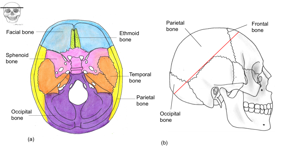
Figure 1. (a) Superior view of cranial base of the neurocranium. (b) Lateral view of skull. Area superior to the red line shows the calvaria of the neurocranium.
viscerocranium
The viscerocranium is situated anterior to the neurocranium and is comprised of 14 irregular bones including 6 pairs of bones:
The viscerocranium develops in the mesenchyme (neural crest cells from which the calvaria of the neurocranium is also derived) or sclerotome (derived from mesoderm) of the 1st and 2nd embryonic pharyngeal arches. The bones of the viscerocranium create what is commonly referred to as the facial skeleton (Figure 2).
Within commonly used texts there is debate as to which bones are included in the facial skeleton (viscerocranium). Some make a distinction between the bones of the skull based on the embryological origins. Other texts include all the bones that can be seen on an anterior view of the skull as part of the . As a result, it is not uncommon to see the frontal, hyoid, parietal and sphenoid bones described as part of the facial skeleton.
- Zygomatic (x2)
- Palatine (x2)
- Nasal (x2)
- Lacrimal (x2)
- Maxilla (x2)
- Inferior nasal concha (x2)
- Mandible
- Ethmoid
The viscerocranium develops in the mesenchyme (neural crest cells from which the calvaria of the neurocranium is also derived) or sclerotome (derived from mesoderm) of the 1st and 2nd embryonic pharyngeal arches. The bones of the viscerocranium create what is commonly referred to as the facial skeleton (Figure 2).
Within commonly used texts there is debate as to which bones are included in the facial skeleton (viscerocranium). Some make a distinction between the bones of the skull based on the embryological origins. Other texts include all the bones that can be seen on an anterior view of the skull as part of the . As a result, it is not uncommon to see the frontal, hyoid, parietal and sphenoid bones described as part of the facial skeleton.
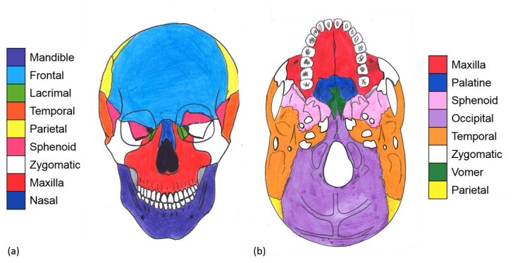
Figure 2. (a) Anterior view of the facial bones of the skull (viscerocranium). (b) Inferior view of the skull base with mandible removed showing the relations of the bones of the viscerocranium and neurocranium.
pop quiz
Check how much of the skull development section you understand by completing the quiz below.
- To begin click on 'Skip step'
- Then click 'Start Quiz'
- Good luck!
Keep attempting the quiz until you get more than 60%. Use the hint button under the questions to view the answer if you are stuck!

