CRANIAL FOSSAE
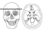
The internal surface of the neurocranium base has 3 depressions which create the bowl shape of the cranial cavity that accommodate the brain. Figure 1 displays the 3 depressions/fossae. The fossae increase in depth from anterior to posterior and are termed the:
- Anterior cranial fossa
- Middle cranial fossa
- Posterior cranial fossa
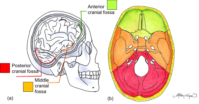
Figure 1. (a) Lateral view of cranial fossae of the adult skull with brain in situ. (b) Superior view of cranial fossae of adult skull with brain removed.
ANTERIOR CRANIAL FOSSA
The anterior fossa is formed from the frontal bone anteriorly, the ethmoid bone in the midline and the body and the lesser wings of the sphenoid bone posteriorly (Figure 2). Two skull foramina located in the anterior fossa:
- Foramen caecum
- Foramen of the cribriform plate
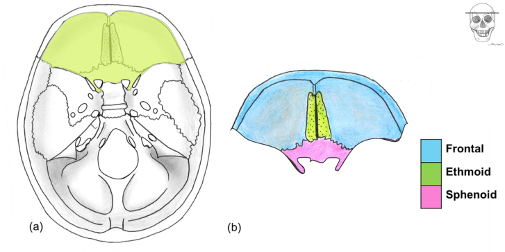
Figure 2. (a) Superior view of the skull base with the anterior cranial fossa highlighted in green. (b) Bones that form the anterior cranial fossa.
Middle cranial fossa
The middle cranial fossa is butterfly shaped and is located posteroinferior to the anterior fossa (Figure 3). Both the greater wings of the sphenoid and temporal bone create the lateral sections of the fossa. The middle cranial fossa contains 6 foramina:
- Optic canal
- Superior orbital fissure
- Foramen rotundum
- Foramen ovale
- Foramen spinosum
- Foramen lacerum
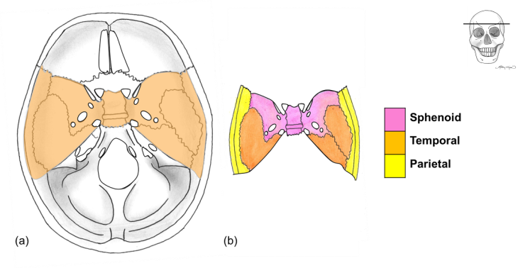
Figure 3. (a) Superior view of the skull base with the middle cranial fossa highlighted in orange. (b) Bones that form the middle cranial fossa.
Posterior Cranial Fossa
The posterior fossa is the largest and deepest of the 3 fossae. The occipital bone is the main contributor to the fossa and the temporal bone forms the antero-lateral boundaries (Figure 4). There are 4 foramina found in the posterior cranial fossa:
- Internal acoustic meatus
- Jugular foramen
- Hypoglossal canal
- Foramen magnum
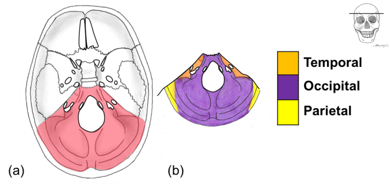
Figure 4. (a) Superior view of the skull base with the posterior cranial fossa highlighted in red. (b) Bones that form the posterior cranial fossa.
POP QUIZ
Check how much of the cranial fossae section you understand by completing the quiz below.
- To begin click on 'Skip step'
- Then click 'Start Quiz'
- Good luck!
Keep attempting the quiz until you get more than 60%. Use the hint button under the questions to view the answer if you are stuck!

